
A surface design that kills bacteria without antibiotics could point to the next stage of medical AM
Researchers at the Indian Institute of Technology Mandi (IIT Mandi) have developed a ceramic coating for 3D-printed bone implants, modeled on the spiny structure of sea urchins. The goal is to solve two chronic problems in bone implants, infection and poor bone integration, through a single surface treatment. The findings were published in the journal Chemical Engineering Journal.
Needle-Like Hydroxyapatite Clusters Physically Damage Bacteria While Promoting Bone Bonding
Treating large bone defects caused by trauma, infection, or tumor removal remains difficult. 3D-printed implants made from polylactic acid (PLA) can be customized to match a patient’s specific bone defect, but PLA’s hydrophobic nature has long made it hard to bond effectively with bone. Bacteria adhering to an implant surface and forming biofilms can also lead to implant failure and additional surgery.
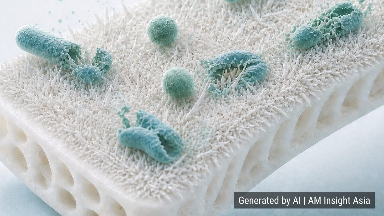
A team led by Dr. Sumit Murab, together with Ankita Negi, Aakash Verma, K.M. Mohammed Sufiyan, and Vedante Mishra, developed a dual-layer coating built around hydroxyapatite, the mineral that makes up most of human bone. The coating process happens in two stages. First, a 3D-printed PLA scaffold is treated with an alkaline solution that activates the surface and creates sites where minerals can deposit. Then, hydrothermal treatment at 90°C forms clusters of needle-like hydroxyapatite crystals resembling the spines of a sea urchin.
This structure does more than mimic a natural shape. The hydroxyapatite itself offers a bone-compatible mineral surface, while the needle-like microstructure physically damages bacteria and hinders their growth. Rather than killing bacteria with drugs, the approach relies on the surface’s own geometry to keep bacteria from taking hold. According to the researchers, this could reduce bacterial colonization without relying on antibiotics or conventional antibacterial chemicals.
Part of a Broader Shift Toward Antibiotic-Free Antibacterial Coatings
Surface modification of bone implants has long been an active field of research. Coatings that slowly release antibiotics, or that rely on silver or copper ions, have dominated the space, drawing their effect from drugs or metal ions. IIT Mandi’s approach differs in that it focuses on a physical antibacterial effect built into the structure itself.
The idea of killing bacteria through shape alone, without drugs, is not science fiction. It belongs to an established line of research. In 2012, scientists reported that the surface of cicada wings is covered in densely packed, nanometer-scale protrusions called nanopillars. When bacteria come into contact with them, their cell membranes stretch across the gaps between pillars and rupture, killing the bacteria. To confirm that this effect was physical rather than chemical, researchers coated cicada wings with a thin layer of gold to alter their surface chemistry. The antibacterial effect held steady, confirming that the shape of the pillars, not their chemistry, was doing the damage. This phenomenon became known as the mechano-bactericidal effect. Similar nanostructures were later found on dragonfly wings, and researchers have since worked to recreate comparable protrusions on artificial materials such as titanium.
The needle-like hydroxyapatite clusters IIT Mandi developed appear to belong to this same lineage. Where the nanopillars on cicada wings stretch and rupture cell membranes, the needle-shaped hydroxyapatite crystals are thought to pierce or deform bacterial membranes on contact, though the exact mechanism has not been confirmed. The details of how this damage occurs remain debated, with no full consensus among researchers, and the effect’s strength varies depending on the bacterial species (gram-positive or gram-negative) and the shape of the protrusions. It is not a universal antibacterial solution.
At the same time, media coverage of the research has described the two-stage coating process and its purpose in detail but has not mentioned specific plans for animal testing, clinical applications, or quantitative data such as bacterial kill rates. The work currently appears to be at the academic publication stage, with real-world clinical validation still ahead.
AM Insight Asia Perspective
At a time when medical AM news tends to be dominated by corporate announcements out of China and the West, basic research from an Indian university making its mark in the field is worth noting. This is not an isolated case for IIT Mandi: the Jaiswal lab at the same institution has previously published antibacterial implant coatings using sugar-functionalized MoS2 nanosheets, and multiple labs at the university have produced sustained output at the intersection of medical materials and AM. India’s presence in this space is worth tracking not only through national AM industrial strategy, but through the depth of its academic research base.
A second point concerns how the category of 3D-printed implants itself is evolving. The value once centered on shape customization, the ability to match an implant to a patient’s specific bone geometry. As surface coating technologies like this one accumulate, implants are shifting toward complex, multifunctional products that combine shape with infection control and bone bonding. In AMIA’s view, product differentiation is shifting away from printer and filament performance and toward post-processing surface treatment. This shift could open new entry points into the medical AM market for materials makers and coating technology providers.
About the Company
IIT Mandi (Indian Institute of Technology Mandi) is a national engineering institute based in Kamand, Himachal Pradesh, in northern India. The Murab Lab (Translational Tissue Engineering Lab), which led this research, is part of the School of Biosciences and Bioengineering and focuses on regenerative materials for orthopedic tissue as well as 3D printing and bioprinting materials.